Truncus Arteriosus
Download this information sheet as a PDF
The aim of this information sheet is to explain what Truncus Arteriosus (or Common Arterial Trunk) is, what effect it will have on a child and how it can be treated.
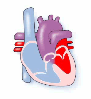
Animation of Truncus Arteriosus
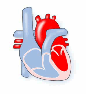
Animation of normal heart
What is Truncus Arteriosus?
When a baby’s heart is developing in the womb, one large blood vessel, the truncus arteriosus, should divide to become the two main arteries: the aorta, and the pulmonary artery.
The aorta should carry red (oxygenated) blood from the heart to the body, and the pulmonary artery should carry blue (deoxygenated) blood to the lungs.
If the aorta and pulmonary artery do not divide, the child is born with a persistent truncus arteriosus or common arterial trunk. This one big artery has one large valve instead of there being pulmonary and aortic valves.
A part of the wall between the left and right ventricles is missing (VSD) so that this single blood vessel receives blue blood from the right side of the heart and red blood from the left side of the heart.
The Truncus Arteriosus divides into the aorta and the pulmonary arteries, which carry this mixture of blood to the lungs and to the body:
- In Type 1 the pulmonary artery branches arise just above the valve.
- Type 2 the pulmonary arteries to the lungs branch separately but close together directly off the truncus.
- Type 3 the pulmonary arteries branch separately and are further apart.
There may be other differences that affect how your child is treated – the VSD may be missing, the valve may not work well and may leak or be narrowed.
The result of Truncus Arteriosus is usually that too much blood is going to the lungs and too little red (oxygenated) blood is reaching the body.
Diagnosis
Truncus Arteriosus can sometimes be seen on a scan during pregnancy.
After birth your baby may have been diagnosed because a heart murmur was heard. The child becomes increasingly blue (cyanosed), because of the blue blood being pumped to the body instead of the lungs.
Due to high pressure in the lung, he or she will develop congestive heart failure as blood flow to the lungs increases. This results in breathlessness, difficulty in feeding and ‘failure to thrive’.
When a heart problem is suspected the tests used can be:
- pulse, blood pressure, temperature, and number of breaths a baby takes a minute
- listening with a stethoscope for changes in the heart sounds
- an oxygen saturation monitor to see how much oxygen is getting into the blood
- a chest x-ray to see the size and position of the heart
- an ECG (electrocardiogram) to check the electrical activity
- an ultrasound scan (echocardiogram) to see how the blood moves through the heart
- checks for chemical balance in blood and urine
- a catheter or Magnetic Resonance Imaging test may be needed.
Treatment
The aim of the treatment is to separate the circulation of the blood so that blue blood goes to the lungs, and red to the body.
Risk of Procedures
There is a risk to your child in the procedures described, but how great that risk is depends on how severe the heart defect is and how well your child is otherwise. The doctors will discuss risks with you in detail before asking you to consent to any of the operations.
Although the time is often very short to take decisions on behalf of a newborn, you will find that later decisions often leave you and your child several months or even years to decide the best time for more treatment.
Surgery
Surgery will be needed before the increased pressure irreparably damages the lungs (pulmonary vascular disease). This will be open heart surgery. The heart will need to be stopped and opened to repair it. This means that a machine will have to take over the job that the heart and lungs normally do – the heart bypass machine.
The usual procedure is to place a donor lung artery (called a homograft or conduit) from the right ventricle to the branch lung arteries and a plastic patch is usually used to close the hole (VSD).
There is no risk of rejecting the donor lung artery, since it is not living blood vessel.
The truncus will now act as the aorta, carrying red blood from the left ventricle to the body. Surgeries will need to be repeated as your child grows.
The more complicated types of Truncus may be treated with a Fontan-style of operation.
How the child is affected
The surgery changes the pressures inside the heart, and it may take your baby a while to adapt to the new ones. It is not uncommon for a child to pick up an infection, such as a chest infection or infected wound while undergoing treatment. Some children react badly to some kinds of medicines.
The kind of surgery needed can sometimes cause a very fast (tachycardia), or very slow (bradycardia) pulse rate, which may need medication to keep it stable.
Children with Truncus Arteriosus often have other health problems, which can include Di George syndrome caused by a genetic defect and results in a small or absent thymus gland which leaves them open to recurrent infection.
Most babies are well, active, and gaining weight a few days after surgery. He or she will have a scar down the middle of the chest, and there may be small scars where drain tubes were used. These fade very rapidly in most children, but they will not go altogether. Smaller scars on the hands and neck usually fade away to nothing.
Unfortunately, because the replacement lung artery or conduit that has been put in doesn’t grow with the child, further surgery will be needed as he or she gets bigger – perhaps another two or three open heart surgeries.
Because the flow of blood is slower than it should be in the conduit, there may be a danger of blood clotting – if this is thought to be a problem in your child’s case, he or she may need to take aspirin or Warfarin.
Aspirin and warfarin have an anticoagulant effect to stop blood cells sticking together. Warfarin does need to be checked by a blood test every few weeks.
At home
While you are at home:
- You, your GP and Health Visitor should have details of your baby’s condition from the heart doctor (paediatric cardiologist). If not, call the hospital at which your baby was treated, ask for the name of the paediatric cardiologist and their telephone number. Call and explain that you need the information to pass on to, for example, your local accident and emergency Department should he or she have a sudden illness.
- You should have the number of a cardiac liaison nurse or outreach nurse to call should you have questions or any fears about your baby’s heart problem.
- You should have the number of a parent support group.
The child will be monitored regularly by a cardiologist.
Some of these problems can occur after surgery or later in life:
- The Truncal valve may need replacement with an artificial valve. If this happens the child may need to take an anticoagulant medicine to stop blood clots forming on the new valve. The anticoagulant effect has to be monitored frequently using a blood test.
- The electrical system of the heart is sometimes damaged, causing a very fast heart beat (tachycardia) or a slow heart beat (bradycardia) to occur. This may be improved with medicine, ablation (a catheter inserted through a vein to stop fast rhythm problems) or a pacemaker.
Evidence and sources of information for this CHF information sheet can be obtained at:
(1) NHS Choices. Congenital Heart Disease Conditions. London: NHS; 2017. Available at:
www.nhs.uk/conditions/congenital-heart-disease/Pages/Types.aspx
(2) World Journal for Pediatric and Congenital Heart SurgeryFirst Published January 25, 2017. Intramural Coronary Artery in Truncus Arteriosus: Importance of Preoperative Echocardiographic Diagnosis and Impact on Surgical Planning. Arshid Mir, MD, et al. Available at:
https://doi.org/10.1177/2150135116682453
About this document:
Published: May 2013
Reviewed: May 2022
To inform CHF of a comment or suggestion, please contact us via info@chfed.org.uk or Tel: 0300 561 0065.